2015-06-03 01:09:59Elisabeth Abrahamian
Working With Arch Pain
Overview
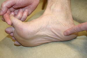
The arch of the foot is made from a complex series of bones tendons and ligaments. According to podiatric practice there is only one important arch of the foot on the medial side. The height of the arch varies and having a flat foot or high arched foot is inherited. The arch acts to dissipate forces and store energy within the foot. Most young children have flat feet and this is because the arch has fatty tissue overlying it. Normally an arch appears after the age of 5-6 years. Even if you remain flat footed this might be normal for you and your family. Just because the arch is low does not mean that any treatment is required. Many world class athletes have flat feet. If the arch is painful or the arch of one foot suddenly drops then investigation and treatment may be required. The arch might drop acutely due to a tear in a tendon or the movement of a bone within the arch. A scan and X Ray may be indicated and treatment ranging from insoles to surgery may be required. If you have a high arched foot this is likely to have been inherited. In extreme form this can cause pain in the ball of the foot and is often associated with a tight Achilles tendon and a tendency to walk on the outside of your foot. In most people there are no symptoms and in others there may pain along the arch and pain on the outside of your ankle. Both these foot types are often mild and require no treatment, but if symptoms occur a full bio-mechanical examination will often reveal a mechanical cause to your pain. Often the treatment may be simple stretches and orthoses.
Causes
The arch of the foot is the concaved, mid-section of the sole. While it only spans an inch or two in most adults, this one small area of the foot bears nearly all of your weight when you walk, and helps to transfer this weight from heel to ball. Just beneath the skin on the sole of the foot, a tough, elastic ligament called the plantar fascia extends from your heel bone to the metatarsal area of the foot. This ligament is designed to bounce gently with the spring of your step, but a number of factors can cause it to become unhealthy. These include. An abnormal walking gait. Vigorous high-impact exercise such as running, playing tennis or basketball. Being overweight. Wearing shoes that slant or cramp any part of the foot. Wearing shoes that have worn down in the heel or sole. A traumatic injury to the foot, including cuts, bruises, strains and fractures. The presence of arthritis or other inflammatory conditions. The normal aging process. In the presence of any of the above factors, the plantar fascia ligament can begin to flex beyond its normal range of motion. Small tears may develop in the tissue and inflammation is commonly present. You may describe your arch pain as sore, sharp, tender, intermittent, constant, burning, tingling or aching. All of these adjectives may be signs that you are experiencing a condition called Plantar Fasciitis.
Symptoms
Intense heel pain, especially first thing in the morning and after a long day. Difficulty walking or standing for long periods without pain. Generally, the sharp pain associated with plantar fasciitis is localized to the heel, but it can spread forward along the arch of the foot and back into the Achilles tendon. While severe cases can result in chronic pain that lasts all day, the most common flare ups occur first thing in the morning, making those first steps out of bed a form of torture, and in the evening after having spent a day on your feet. Overpronation (a foot that naturally turns too far inward), high arches, and flat feet (fallen arches) can all cause similar arch pain. In these cases, however, the pain is more likely to continue throughout the day rather than being worst in the morning.
Diagnosis
The adult acquired flatfoot, secondary to posterior tibial tendon dysfunction, is diagnosed in a number of ways with no single test proven to be totally reliable. The most accurate diagnosis is made by a skilled clinician utilizing observation and hands on evaluation of the foot and ankle. Observation of the foot in a walking examination is most reliable. The affected foot appears more pronated and deformed compared to the unaffected foot. Muscle testing will show a strength deficit. An easy test to perform in the office is the single foot raise.
Non Surgical Treatment
The treatment is to put an arch support under the foot immediately to prevent the arch from collapsing and the plantar fascia from stretching. Also, put an arch support in your slippers and wear them as soon as you rise. Even a few steps barefoot without support can stretch the plantar fascia. Arch supports usually relieve pain within a few days. To head off arch pain, begin an exercise routine slowly, take off any excess weight and wear arch supports in your athletic shoes. Arch pain commonly smolders for months because people do not take the proper precautions. Continuing to do weight-bearing exercises will perpetuate the pain. While the foot is recovering, swim or do water workouts. Or work the upper body only. Some people are able to use a stationary bicycle by placing only the front part of the foot on the pedals.
Surgical Treatment
Foot surgery is difficult, especially when large amounts of deformity correction are needed. The ability to bring the foot into a new position may not be lasting, even if everything looks perfect in the operating room. The goal is to provide improved position and function of the foot and ankle. In some patients with very severe deformity, the goal is a foot that functions well in a brace. There are complications that relate to surgery in general. These include the risks associated with anesthesia, infection, damage to nerves and blood vessels, and bleeding or blood clots. Particular complications associated with cavus foot surgery include incomplete correction of deformity, return of deformity and incomplete fusion.
Prevention
The best method for preventing plantar fasciitis is stretching. The plantar fascia can be stretched by grabbing the toes, pulling the foot upward and holding for 15 seconds. To stretch the calf muscles, place hands on a wall and drop affected leg back into a lunge step while keeping the heel of the back leg down. Keep the back knee straight for one stretch and then bend the knee slightly to stretch a deeper muscle in the calf. Hold stretch for 15 seconds and repeat three times.
The arch of the foot is made from a complex series of bones tendons and ligaments. According to podiatric practice there is only one important arch of the foot on the medial side. The height of the arch varies and having a flat foot or high arched foot is inherited. The arch acts to dissipate forces and store energy within the foot. Most young children have flat feet and this is because the arch has fatty tissue overlying it. Normally an arch appears after the age of 5-6 years. Even if you remain flat footed this might be normal for you and your family. Just because the arch is low does not mean that any treatment is required. Many world class athletes have flat feet. If the arch is painful or the arch of one foot suddenly drops then investigation and treatment may be required. The arch might drop acutely due to a tear in a tendon or the movement of a bone within the arch. A scan and X Ray may be indicated and treatment ranging from insoles to surgery may be required. If you have a high arched foot this is likely to have been inherited. In extreme form this can cause pain in the ball of the foot and is often associated with a tight Achilles tendon and a tendency to walk on the outside of your foot. In most people there are no symptoms and in others there may pain along the arch and pain on the outside of your ankle. Both these foot types are often mild and require no treatment, but if symptoms occur a full bio-mechanical examination will often reveal a mechanical cause to your pain. Often the treatment may be simple stretches and orthoses.

Causes
The arch of the foot is the concaved, mid-section of the sole. While it only spans an inch or two in most adults, this one small area of the foot bears nearly all of your weight when you walk, and helps to transfer this weight from heel to ball. Just beneath the skin on the sole of the foot, a tough, elastic ligament called the plantar fascia extends from your heel bone to the metatarsal area of the foot. This ligament is designed to bounce gently with the spring of your step, but a number of factors can cause it to become unhealthy. These include. An abnormal walking gait. Vigorous high-impact exercise such as running, playing tennis or basketball. Being overweight. Wearing shoes that slant or cramp any part of the foot. Wearing shoes that have worn down in the heel or sole. A traumatic injury to the foot, including cuts, bruises, strains and fractures. The presence of arthritis or other inflammatory conditions. The normal aging process. In the presence of any of the above factors, the plantar fascia ligament can begin to flex beyond its normal range of motion. Small tears may develop in the tissue and inflammation is commonly present. You may describe your arch pain as sore, sharp, tender, intermittent, constant, burning, tingling or aching. All of these adjectives may be signs that you are experiencing a condition called Plantar Fasciitis.
Symptoms
Intense heel pain, especially first thing in the morning and after a long day. Difficulty walking or standing for long periods without pain. Generally, the sharp pain associated with plantar fasciitis is localized to the heel, but it can spread forward along the arch of the foot and back into the Achilles tendon. While severe cases can result in chronic pain that lasts all day, the most common flare ups occur first thing in the morning, making those first steps out of bed a form of torture, and in the evening after having spent a day on your feet. Overpronation (a foot that naturally turns too far inward), high arches, and flat feet (fallen arches) can all cause similar arch pain. In these cases, however, the pain is more likely to continue throughout the day rather than being worst in the morning.
Diagnosis
The adult acquired flatfoot, secondary to posterior tibial tendon dysfunction, is diagnosed in a number of ways with no single test proven to be totally reliable. The most accurate diagnosis is made by a skilled clinician utilizing observation and hands on evaluation of the foot and ankle. Observation of the foot in a walking examination is most reliable. The affected foot appears more pronated and deformed compared to the unaffected foot. Muscle testing will show a strength deficit. An easy test to perform in the office is the single foot raise.
Non Surgical Treatment
The treatment is to put an arch support under the foot immediately to prevent the arch from collapsing and the plantar fascia from stretching. Also, put an arch support in your slippers and wear them as soon as you rise. Even a few steps barefoot without support can stretch the plantar fascia. Arch supports usually relieve pain within a few days. To head off arch pain, begin an exercise routine slowly, take off any excess weight and wear arch supports in your athletic shoes. Arch pain commonly smolders for months because people do not take the proper precautions. Continuing to do weight-bearing exercises will perpetuate the pain. While the foot is recovering, swim or do water workouts. Or work the upper body only. Some people are able to use a stationary bicycle by placing only the front part of the foot on the pedals.

Surgical Treatment
Foot surgery is difficult, especially when large amounts of deformity correction are needed. The ability to bring the foot into a new position may not be lasting, even if everything looks perfect in the operating room. The goal is to provide improved position and function of the foot and ankle. In some patients with very severe deformity, the goal is a foot that functions well in a brace. There are complications that relate to surgery in general. These include the risks associated with anesthesia, infection, damage to nerves and blood vessels, and bleeding or blood clots. Particular complications associated with cavus foot surgery include incomplete correction of deformity, return of deformity and incomplete fusion.
Prevention
The best method for preventing plantar fasciitis is stretching. The plantar fascia can be stretched by grabbing the toes, pulling the foot upward and holding for 15 seconds. To stretch the calf muscles, place hands on a wall and drop affected leg back into a lunge step while keeping the heel of the back leg down. Keep the back knee straight for one stretch and then bend the knee slightly to stretch a deeper muscle in the calf. Hold stretch for 15 seconds and repeat three times.